TREE LIFE
March 1997
ANNUAL SUBS – NOW DUE
Happily it has not been necessary to increase our subs this year. Please send in your $40 promptly with the invoice which can be found in the body of this Tree Life.
MASHONALAND CALENDAR
Tuesday 4th March: Botanic Garden Walk. We will meet Tom in the car park at 4.45 for 5 p.m. and continue with the fascinating topic of creepers. This will be Part 2 as last month’s walk was also rained-out. There will be a guard for the cars.
Sunday 16th March: There must be some interesting riverine vegetation along the Shavanhohwe River and we will start the walk where the bridge spans the river on the Mutoko Road, and work our way along the banks. There will be a guard for the cars.
Saturday 22nd March: Mark’s Botanic Walk will take place at Domboshawa (planned for January but rained out). The entrance fee is $3 each and we will meet at 2.30 p.m. in the car park.
Thursday 27 to Monday 31 March: – Easter. To Tuli.
Tuesday 1st April: Botanic Garden Walk.
MATABELELAND CALENDAR
Sunday 2nd March: To Bra
Monday 10th March: – Study Session. Urban trails in Circular Drive area. Meeting at 5-5.15 p.m. These sessions will take place on the second Monday of every month.
Sunday 6th April: To Nyamandhlovu to a farm run by Sandy and Philip Palmer.
VISIT BY THE MATABELELAND BRANCH TO LOCHVIEW: THE HOME OF CHRIS AND DICK STEVENS.
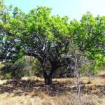
The good rains this season have produced a profusion of exuberant growth in grasses, shrubs, climbers and trees. In fine sunny weather we explored two small areas the first a densely wooded ironstone-like ridge through and around which Chris had cut paths to facilitate access.
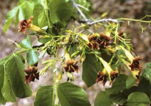
Markhamia zanzibarica. Photo: Darrel Plowes. Source: Flora of Zimbabwe
A large Ficus thonningii stood sentinel with roots pouring over the rock face at the entrance to our route-way. Ehretia rigida bedecked with orange berries was cause for discussion followed by several Markhamia zanzibarica with rich claret-coloured Bignonia flowers tucked under the foliage. Numerous Clerodendrum glabrum were in flower. The thick flowering grasses were interspersed with beautiful profusely blooming Tephrosia rhodesiaca and baby zinnias. Cadaba termitaria not immediately recognised took time to identify, as did Acacia gerrardii. Several species were in fruit including Albizia amara, Peltophorum africanum, Flueggea virosa, Acacia nilotica, Combretum hereroense and Grewia bicolor, Grewia flavescens and Grewia monticola. It was exciting to find Cordia ovalis – not listed on our Bulawayo District card, though we have seen it on the Shashi. In the tangled mass, we were able to see Acacia schweinfurthii in flower, Euphorbia tiru-calli, Dovyalis caffra and Dovyalis zeyheri, Ficus ingens, a curtain of Gymnema sylvestre patterned with delicate little yellow flowers (an Asclepiadaceae climber) dozens of Ximenia americana and, not so common in our usual haunts, Grewia flava. As we left our little jungle we disturbed an owl, possibly two.
Tea was taken in the shade of a large Belhambra, Phytolacca dioica, and the vast root mass providing convenient seating.
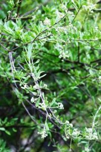
Boscia angustifolia, Photo: Bart Wursten. Source: Flora of Zimbabwe
We then walked round the kopjie behind the homestead. A fruiting Berchemia zeyheri was a mass of delicious little red berries. Next to it in line, although planted, were three Schrebera alata and at the crest of the hill a Boscia angustifolia, not so frequently seen on our outings.
A Grewia flavescens var. olukondae was in fruit and an unidentified Barleria sp. shrub was in flower.
All too soon the morning was over, the time had come to bid farewell to Chris. Our thanks to her and to Dick for inviting us to share their little wilderness area which yielded 53 tree species and I’m sure held much of interest which we missed.
-Margaret McCausland.
To add to Margaret’s account of our visit, it would appear that Cordia ovalis has been combined ‘perhaps temporarily’ with Cordia sinensis in a recent revision of the Boraginaceae by E.S. Martins in Flora Zambesiaca Vol. 7 Part 4 1990. Other name changes in the genus Cordia are: ¬ Cordia abyssinica now becomes Cordia africana. Lam., Tab. Encycl. M’eth. Bot. 1:420 (1792) Type from Ethiopia.
Cordia mukuensis Taton in Bull. Jard. Bot. Nat Belg. 41: 258 (1971) – not previously listed as occurring in Zimbabwe.
Cordia monoica R oxb., P Coromandel 1: 43, 58 (1795) not previously listed for Zimbabwe – this species includes Cordia ovalis Hochst ex A. DC., Prodr. 9; 479 (1845).
Cordia sinensis lam Tab. Encycl. M’eth. Bot. 1: 423 (1792). This includes Cordia ovalis sensu Palmer & Pitman. Trees of Southern Afr. 3: 1939 (1973).
Without going into further detail, it would appear that what we have known as Cordia ovalis is now Cordia sinensis (or possibly).
The author E. S. Martins did mention that more comprehensive studies were lacking, but nevertheless he had decided to include several species in Cordia sinensis at least provisionally.
So until further work is done Cordia ovalis is Cordia sinensis.
-A. Ellert.
MARKWE CAVE, 19 JANUARY 1997
It was a dark and stormy night, and the morning wasn’t much better either. However, around 67 souls decided to brave the cold, overcast conditions and were rewarded with one brief shower and a beautiful day. Tree Soc. was privileged to be joined by Prehistory Society members and the day started with a brief lesson on the graves and paintings of Markwe Cave by Rob Burrett.
The graves are those of headmen or chiefs from the last 500 years (dated by the stone walling). The bodies were placed in natural hollows in the stone and then walled in. Remains of clay pots around the graves were probably for libations to be paid to the ancestral spirits. Most of the walls were moulded with clay, some quite recently. The paintings at the site date from between 10000 to 500 years ago and the art is not meant to be realistic but rather symbolic, displaying religious and social concepts. A large elephant was outlined and the central area had faded and previous paintings were visible underneath. Such painting layers can represent associations between pictures. Rob also pointed out the facing male and female figures in leg-bent posture, the figure in a tranced state, the obese “mother goddess”, the mythological looking antelope-headed guineafowl and others. All-in-all extremely interesting even though trees were not mentioned!
Whilst the brave and the foolish decided to scale the slippery slope up to see the cave, others milled aimlessly around taking in the scenery and marvelling at the many colourful mushrooms and fungi (is there no scope for a Fungus Soc.?) These were to feature throughout the marshy woodland with many Cantharellus species (gold, orange, red or brown caps which are depressed in the centre and with wavy margins and only weakly inter-veined gills) and some beautiful (and poisonous) Amanita specimens.
Many of these were trodden on or uprooted as we strolled under the large Brachystegia spiciformis mixed with Erythrina abyssinica, Heteropyxis dehniae, Clerodendrum wildii, Erythroxylum emarginatum, Rhus longipes, Vitex payos, Ximenia caffra (no sour plum fruit to be seen), Tetradenia riparia (ginger bush) and, of course, Fagaropsis angolensis. The latter had the characteristic opalescent glands around the leaf margins. Also visible were Pterocarpus angolensis and Cussonia natalensis.
When we had Strychnos cocculoides (paired hooked spines but with corky bark), Combretum molle (velvet-leafed Combretum). Julbernardia globiflora (Mnondo – often mistaken for Msasa but has more pairs of leaflets and the last pair are smaller than the rest). Grewia monticola (asymmetric base to leaf, 3 veins from base, lighter under surface of leaf – don’t mistake it for Ziziphus; the leaf is still a bit sandpapery and there are no thorns). Grewia decemovulata (a shrub with small yellow flowers) and Ziziphus mucronata (Buffalo thorn) pointed out to us; most of us had half our attention on the ground trying to dodge the springhare and aardvark holes. A gall-stricken Pseudolachnostylis maprouneifolia attracted my attention. There was also an opportunity to compare Maytenus heterophylla and Maytenus senegalensis together and I was struck by the profusion of leaves on the former, not just the fact that it has leaves of many sizes and no pink petioles. Continuing to wind along the base of the kopjie Ochna schweinfurthiana in fruit (4 red carpels opened out to expose the black seeds) and a variable Flacourtia (almost hibiscus-shaped leaves and long spines) caught the eye. However, stomachs were starting to grumble and Mundulea sericea, Terminalia sericea, Ozoroa insignis, Schrebera alata, Peltophorum africanum, Senna singueana, Albizia antunesiana, Azanza garckeana, Lannea edulis, Elephantorrhiza goetzei, Erythrococca trichogyne, Faurea saligna, Hymenodictyon floribundum and Parinari curatellifolia got as much notice from me as they have space in this write-up! I had even less time for the Jacaranda mimosifolia but managed to stop and look at the hairy caterpillars starting to pupate on the prickly pear. (The spines of the caterpillar are very irritating as Mary Toet can witness to). I also had to crush the leaves of Steganotaenia araliacea to breathe in the fresh carroty smell. With lunch under digestion it was time for afternoon exercise – the assault on the hill. Led past Cussonia arborea & Tarenna neurophylla, Rhoicissus tridentata, Solanum sp., Combretum zeyheri and others our wise leaders showed us that it only requires positive psychology to climb a vertical granite wall when there is a much easier route round the other side. Yet, after much huffing and puffing, we were rewarded with a glorious view of the surrounding countryside, not to mention the Gloriosa superba (flame lilies) growing amongst the resurrection plants and Xerophyta. Ficus sycomorus (thank you Karl for correcting Andy) and Ficus thonningii were present up the top where the rocks were decorated by patterns of yellow, orange, green and white lichens. Walking past the ‘swimming pool’ (I’d leave it to the tadpoles personally) various groups started to wander across the granite dome either to wend their way back to the cars or to look at isolated clumps of trees. Landed with having to write this article I thought I’d better see what else could be found.
Commiphora africana, Commiphora marlothii, Dovyalis zeyheri, Grewia flavescens (square stem), Lannea discolor (not datcolor!). Pouzolzia mixta (soap nettle), Pterocarpus rotundifolius (with yellow flowers). And Dombeya rotundifolia shielded a patch of heavily spined Pterolobium in which flourished two Synadenium africanum – the dead-man’s tree, a sight so rare that it doesn’t even feature on the tree cards! This Euphorbiaceae also has irritant white latex and is supposed to have the bones of dead animals lying around its base since it so poisonous. I failed to see any but was not surprised. Hidden amongst the miniature forest it seemed an appropriate moment to swap jokes. Since us men were outnumbered five to three we had to just accept that “jokes about blondes are short so that men can understand them” and “the way to get a man to commit suicide is to get him to jump from his ego to his IQ”. Reassembling our split sides we headed back down the kopjie for fluid replenishment and a final chat. Overall a wonderful outing in a special area and our thanks to the Swansons for allowing us to spend the day there.
-Douglas Ball.
IN SEARCH OF TRICERATELLA
Triceratella is a genus in the family Commelinaceae. There is only one species, Triceratella drummondii, a small yellow-flowered annual, and this species has been found only in one place in the world, near Chiturapadzi, east of Beitbridge, in southern Zimbabwe. It was first found by Bob Drummond in May 1958 and the genus and species were both described by Dr Brenan at Kew in Volume 1 of Kirkia (1960-1961).
Bob saw it again at the same place 10 years later but it has never been seen since. On 16th February 1997, a party of 7 set out to try and re-find it. The main reason was the presence of Bob Faden, who is a botanist at the Smithsonian in Washington, an authority on the Commelinaceae, who is currently writing the accounts for Flora Zambesiaca and Flora of Tropical East Africa. Also present were Bob Drummond, Maureen Silva-Jones, Jonathan Timberlake, Anthony Mapaura, Peter Winter and myself.
Chiturapadzi was easily reached over 80km of good but corrugated dirt road. Then the search began, continuing for the rest of the morning and the afternoon, but, sadly with no success. The area was dry (very dry compared to what we’ve experienced in Harare lately!) but many other tiny annuals had appeared – but no sign even of an immature Triceratella. However, a number of other exciting plants were seen, including Albizia forbesii, Grewia hornbyi and Ptaeroxylon obliquum.
Bob Faden did not have a totally wasted trip as we found (at other places) 3 other rarely recorded Commelinaceae; namely: Aneilema schlechteri, Aneilema indehiscens and Commelina kotschyi.
It was also a pleasure to see the Save River flowing vigorously across its whole 400m width on the road between Chiredzi and Chisumbanje.
-MA HYDE.
PLANT GALLS
Galls, abnormal growths on plants, have fascinated botanists for many years and even Charles Darwin discussed them as a possible contradiction to his theory of evolution by natural selection. Whilst certain general aspects are known about them, much of the finer detail about how they come about and the relationship between gall-former and plant host are still the subject of some speculation. The treatise by Mani (1964) remains the basis of much of the knowledge of galls today.
What is a gall?
Galls are cells, tissues or organs which have developed abnormally by cell enlargement (hypertrophy) and cell proliferation (hyperplasia) and are caused by the action of biologic organisms such as bacteria, fungi, nematodes, mites and insects.
However, the definition of a gall is not always quite so clear-cut particularly since there are many gradations between what is normal and what is grossly abnormal. For example, some scientists may classify all abnormalities of a leaf caused by a fungus as a gall whereas the stricter definition would require there to be evidence of excess growth i.e. swelling with the plant playing an active role rather than a passive one. Excessive growths can also be caused by mechanical injury and non-biological chemicals but these are not generally considered as galls. Galls may affect any part of a plant including the vegetative parts, reproductive organs and root systems as well as any fruits. Usually they are a direct consequence of the attack by the gall¬-former but in some cases the gall may form at a site distant from the parasite. In such cases the tissue of the gall usually shows normal histology under the microscope (organoid galls) e.g. flower abnormalities in Scabiosa columbaria due to attack of the roots. Organoid galls are usually caused by fungi, mites and aphids. The cells of histioid galls on the other hand differ from those of the organ on which they develop and no normal organ or function can be discerned. This is comparable to the abnormal cell growth on human cancers and, similarly, the changes can be less marked (benign) or extreme (malignant).
Why and how are galls formed?
There is still some debate about whether galls represent a simple parasitic relationship between the gall-former and the plant with the former deriving all the benefit i.e. shelter, nourishment, dispersal to the detriment of the host i.e. loss of nutrients, altered growth patterns, premature decay. There are examples of where the plant itself benefits such as the nitrogen-fixing nodule galls on Leguminosae but in most of these cases the benefit is rather indirect. A logical argument is also that the gall is a simple result of a protective mechanism against the parasite in an attempt to limit the attack and prevent death. It can further be argued that within this interplay there has been mutual adaptation between host and parasite over the years reaching the present state of specialisation. Under these discussions, galls can be due to either parasites or symbiotic organisms.
The discussion on why galls form requires an understanding of how they form but this area is far from being clearly understood. In some cases it may be that the gall-former produces plant hormone analogues which stimulate growth or particular growth patterns, or the parasite is able to mobilise plant-produced hormones. It may also be true that the altered growth may be due to production of cytokines by the plant in response to the attack. In considering these issues one needs to bear in mind the situation of galls produced by insects (Diptera and Hymenoptera) which are usually of a particular growth on a particular part of a particular plant.
Examples of galls.
Galls can be caused by bacteria, fungi, nematodes, mites as well as insects. Viral attack does not seem to commonly give rise to galls.
Galls caused by bacteria.
Agrobacterium tumefaciens causes uncontrolled proliferation of cells at the top of root systems i.e. crown gall, in most hardwoods although there are many species which are not recorded as being hosts. The bacterium enters the tissues through a wound, stimulates increased DNA replication and cell growth and is then sloughed off with external cells back into the soil. However, the gall continues to grow and may even spread to other parts of the plant.
Galls caused by nematodes.
Nematodes almost invariably cause root galls although the attack of the roots can result in organoid galls elsewhere on the plant. The root-knot nematode (Meloidogyne spp.) affects over 1500 species of plants including about 100 species of trees both conifers and hardwoods. The female feeding on the root causes giant cells to form in the root: Cell walls dissolve, nuclear division is increased and the cells enlarge. The female nematode is an endoparasite and lives within the gall feeding on the few giant cells formed. Eggs are extruded together in a sac-like structure on the surface of the gall and the young hatch and may penetrate the root in the same general area.
Some nematodes transmit plant viruses and are important in the transmission of plant disease.
Galls caused by fungi.
Many rust diseases caused by fungi can give rise to gall formation on conifers in particular but also on other trees. Examples are the fusiform rust on southern US pines (caused by Cronartium fusiforme), pine-oak rust (Cronartium quercuum), pine-pine rust (Endocronartium harkenssii) and cedar-apple rust (Gymnosporangium spp.)
Galls caused by mites.
Mites (Acarina) are small eight-legged relatives of spiders which are able to produce much cellular hypertrophy due to their ability to attack individual cells. Some of the galls on inflorescences can develop into witches-brooms i.e. multiple branching. This effect can however also be caused by other sap suckers and fungi.
Galls caused by insects.
The galls caused by insects are numerous. Often the insect lays its eggs within the plant stimulating a gall which then provides nutrition to the grub until it is sufficiently grown. Some of the galls actually have intricately designed hatches or a dehiscent character for dispersal of the inhabitant(s). The galls of Hymenoptera are of the most specialised. The chalcid gall on Eucalyptus sieberiana is noted for its agglomeration of oval, sessile swellings which contain elongated larval chambers.
Zimbabwean Tree Galls
The galls on Zimbabwe’s trees and other plants are not well documented. Some of the more numerous or obvious include the small hairy disc-like scales on the underside of leaves of Parinari curatellifolia which can apparently develop into grey, pointed chambers. Combretum molle galls are large spherical, rugiform (hairy or rug-like) creations usually around leaf axils while those of Terminalia sericea are ovoid, woody and affect the growing shoot of branches.
Conclusion
Galls are a fascinating and mysterious aspect of the interaction of plants with their environment. Further study of the galls and gall-formers of our local vegetation will help to gain a fuller understanding of the interrelationships which exist in our native woodlands and the effect of man on them.
References
Mani MS (1964). The ecology of plant galls. Junk: The Hague
Manion PD (1981) Tree disease concepts. Prentice-hall: Enmglewood Cliffs.
Macnaughton SJ (1983) Physiological and ecological implications of hervivory. In: Lange OL, Nobel PS, Osmond CB, Ziegler H (eds.)
Physiological Plant Ecology Ill: responses to the chemical and biological environment. Springer¬-Verlag: Berlin.
-Douglas Ball.